Real time three dimensional ultrasound scans can almost replace diagnostic hysteroscopy
by Dimitrios Dovas, last updated 17 Feb 2016,
1 min read
It is well known from medical studies that conditions which distort the anatomy of the endometrial cavity can reduce pregnancy rates following IVF treatment. 3D/4D ultrasound scans give us the opportunity to study in detail the endometrial cavity for structural abnormalities or masses that could theoretically affect implantation (polyps, fibroids).
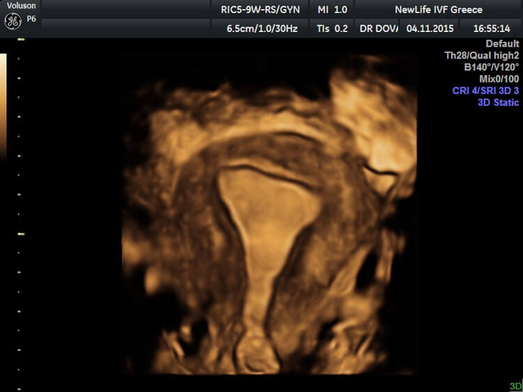
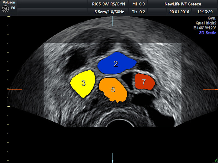
The introduction of this technology reduces the need of invasive diagnostic methods like hysteroscopy. Additionally 3D/4D ultrasound can help us count and measure quickly and accurately the developing follicles during ovarian stimulation with the use of specific software called Sono AVC.
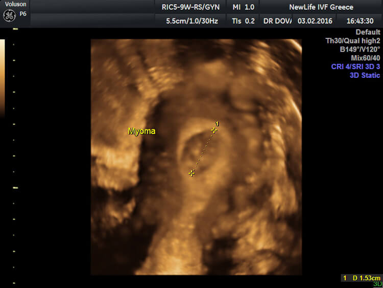
Here at Newlife IVF Greece we offer comprehensive ultrasound scanning investigations not only to monitor response to treatment but also to assess in a non invasive and accurate way the receptivity of the endometrium. Apart from the 3D reconstruction of the endometrial cavity we assess the blood supply towards the uterus and also the vascularity of the endometrium prior to the embryo transfer in order to confirm that we transfer your embryos at the best possible uterine environment.

Dimitrios Dovas, MD, DFFP
Dimitrios is the Clinical Director of Newlife Center of Reproductive Medicine, in Thessaloniki Greece.


