What is the relationship between uterine abnormalities and infertility?
by Newlife IVF Team, last updated 12 Jun 2019,
3 min read
Uterine abnormalities can be congenital - something you are born with - or they can develop over time.
Case 1: Congenital malformations
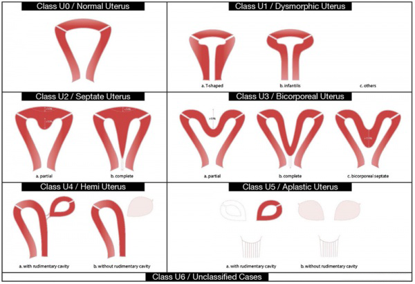
When it comes to congenital malformations are also known as Mullerian anomalies. These abnormalities arise from abnormal development of the uterus, fallopian tubes or cervix. The uterus in early fetal development starts out as two separate parts placed in the upper abdomen. Each part migrates down in the pelvis and joins together to fuse with what will eventually become the vagina. The uterus at that point is still solid and it takes several days to take its final form that in normal cases it has a triangular shape.
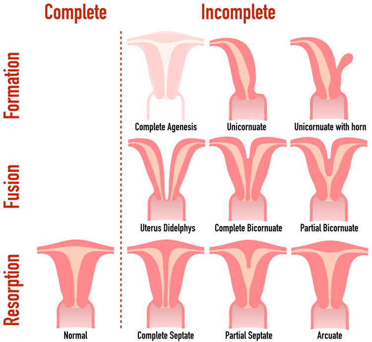
- Septate uterus – is a condition where the middle wall that joins the two uterine cavities fails to completely reabsorb. In this way, there is a single uterine cavity with a defect remaining at the top. This defect can vary in size from very small to one that comes all the way down to the cervix – completely dividing the uterine cavity in two.
- Bicornuate uterus – when the two uterine cavities join only at the bottom (i.e. at the cervix). The upper portions of the uterus are clearly separate. In this situation, there can either be one cervix or two cervices
- Unicornuate uterus – when one side of the uterus does not develop at all, so the ultimate uterus is about half the normal size
- Uterine didelphys – when the separate parts of the uterus don’t join at all, resulting in two separate uterine cavities and two cervices
Each of these abnormalities is associated with both infertility and recurrent pregnancy loss. Only a septate uterus can be surgically repaired, and clinical data suggest that repair results in both significantly higher pregnancy rates and significantly lower miscarriage rates.
Case 2: Abnormalities developed over time
When it comes to abnormalities that have developed over time we imply the formation of:
- a polyp (endometrial)
- uterine fibroids
- uterine adhesions (scarring in the uterine cavity), Asherman’s Syndrome, etc.
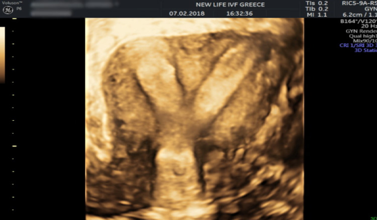
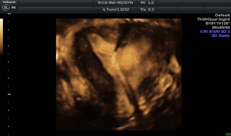
These uterine abnormalities sometimes cause symptoms such as irregular bleeding, heavy periods, pain during menstruation, pain during sexual intercourse, and even frequent urination or constipation. Other times there are no symptoms at all, and these uterine abnormalities are found on a routine ultrasound. Imaging such as a 3D ultrasound or hysterosalpingogram (HSG) can identify these uterine abnormalities.
The application of 3D ultrasound (3DUS) with increased spatial awareness in the last decade of the 20th century has enabled a detailed assessment of uterine morphology. One of the most useful scan planes obtained on 3D ultrasound is the coronal view of the uterus, which is usually not obtainable on 2D ultrasound. Therefore, this view is a valuable problem-solving tool that assists in distinguishing between various uterine abnormalities, including bicornuate, septate, unicornuate and didelphys uterus. Data gaining time is short, and images can be stored for later evaluation and analyzed as many times as necessary. Below there are some images of uterine abnormalities that were detected in our patients during the uterus evaluation scan and the result after the surgical correction of the abnormality.